Laser Scanning Microscopy (LSM)
Short description:
Laser scanning microscopy (LSM) represents a technique which allows assessment of multiple parameters in hydrated samples at the micro-scale and in three dimensions. Laser scanning microscopy is employed for visualization, analysis and quantification. The technique is mainly used for examination of bacteria (size ≈ 1 μm) associated with interfaces and the microbial communities developing over time forming aggregates and biofilms (≈ 150 μm thick). The laser scanning microscopy facility at the UFZ location in Magdeburg is established within the Department of River Ecology. Supervision, support and control of the LSM instrumentation is provided by the research group “Microbiology of Interfaces”. The 3 instruments include: 1) confocal laser scanning microscopy (CLSM) with traditional lasers and multi-photon option; 2) confocal laser scanning microscopy (CLSM) with white laser, lifetime imaging (FLIM) and fluorescence correlation spectroscopy (FCS); and 3) nanoscopy based on ground state depletion (GSD) with 3 laser sources.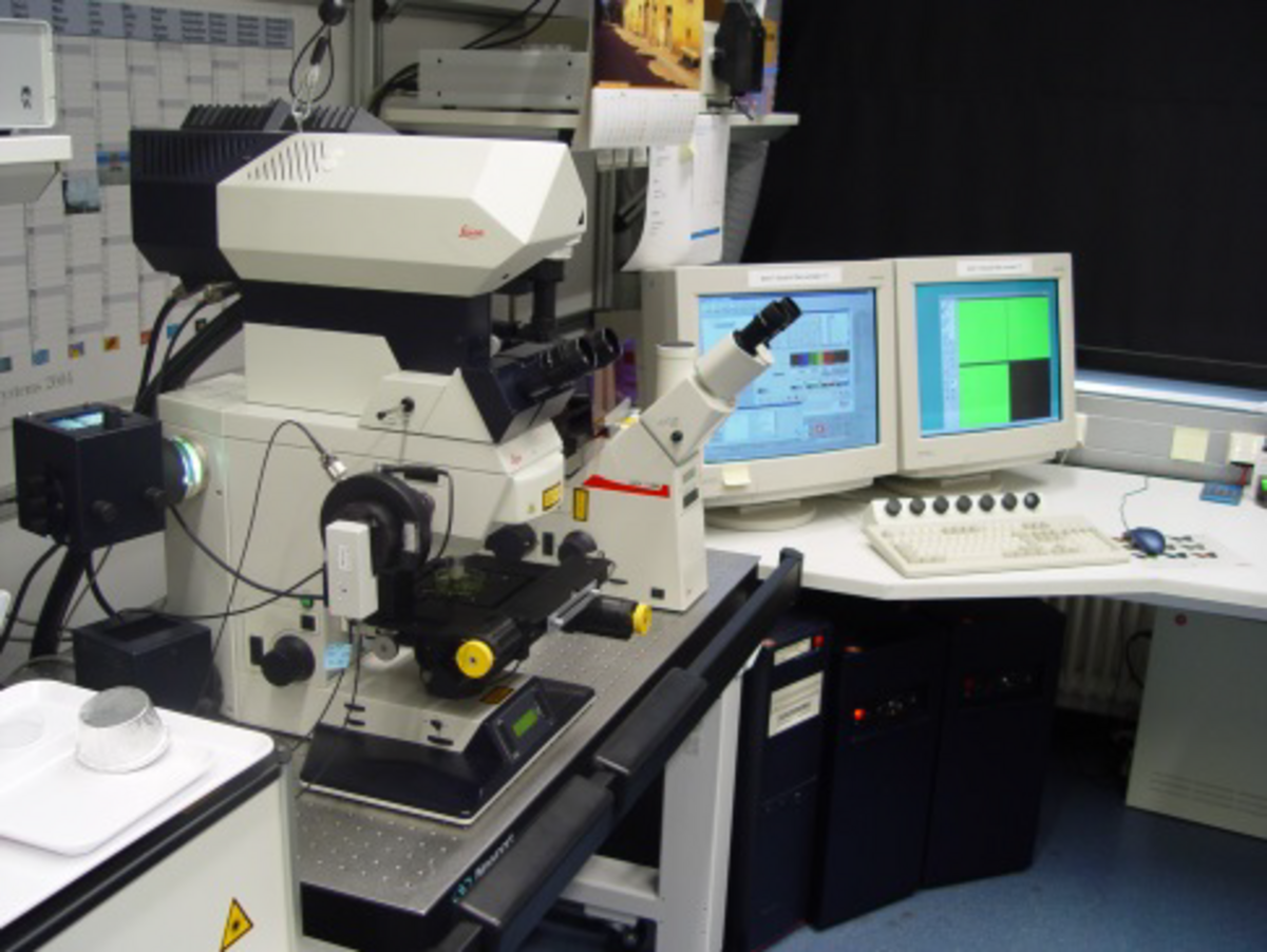
Major research issues/sites:
Conventional light microscopy is limited to samples which are very thin (1-5 μm). In contrast, laser scanning microscopy is a technique that allows assessment of multiple parameters in hydrated samples at the micro-scale and in three dimensions. Laser scanning microscopy is used for visualization, analysis and quantification. Several laser lines can be selected for excitation of fluorochromes specific for different biochemical features. Their emission signals are recorded in separate channels and combined in a 3d multichannel image data set. The technique is mainly used for examination of bacteria (size ≈ 1 μm) associated with interfaces and the microbial communities developing over time forming aggregates and biofilms (≈ 150 μm thick).
Laser scanning microscopy at UFZ started in 1994 after realization that this innovative technique will revolutionize imaging of hydrated (micro-)biological samples. LSM can be used for analysis and understanding of biological systems which cannot be obtained in any other way. It can establish a link between genomic information and function. LSM may be employed to follow the changes in (micro-)biological cell function and processes with time and environment. The 3-dimensional, multi-channel LSM datasets may represent raw data for quantification and modelling.
Main LSM applications are in the field of “structure and function of microbial biofilm systems” especially in the aquatic environment. Special emphasis is put on the composition of the biofilm matrix. Nevertheless the LSM technique is useful in many other biological research fields.
The microbiological issues, applications and expertise are reflected in several invited review articles:
Lawrence JR, Wolfaardt G, Neu TR (1998) The study of microbial biofilms by confocal laser scanning microscopy. In: Wilkinson MHF, Shut F (eds) Digital image analysis of microbes. John Wiley, Chichester, pp 431-465
Neu TR, Lawrence JR (1999) In situ characterization of extracellular polymeric substances (EPS) in biofilm systems. In: Wingender J, Neu TR, Flemming H-C (eds) Microbial extracellular polymeric substances. Springer Verlag, Berlin, pp 21-47
Neu TR (2000) Confocal laser scanning microscopy (CLSM) of biofilms. In: Flemming H-C, Szewzyk U, Griebe T (eds) Biofilms - investigative methods and applications. Springer, Berlin, pp 211-224
Lawrence JR, Korber DR, Wolfaardt GM, Caldwell DE, Neu TR (2002) Analytical imaging and microscopy techniques. In: Hurst CJ, Crawford RL, Knudsen GR, McInerney MJ, Stetzenbach LD (eds) Manual of environmental microbiology, American Society for Microbiology, Washington, pp 39-61
Neu TR, Lawrence JR (2002) Laser scanning microscopy in combination with fluorescence techniques for biofilm study. In: Bitton G (ed) The encyclopedia of environmental microbiology. Volume 4. John Wiley & Sons, New York, pp 1772-1788
Lawrence JR, Hitchcock AP, Leppard GG, Neu TR (2005) Mapping biopolymer distributions in microbial communities. In: Droppo I, Leppard GG, Millard M, Liss SN (eds) Flocculation in natural and engineered environmental processes. CRC Press, Boca Raton, pp 121-141
Palmer Jr. RJ, Haagensen J, Neu TR, Sternberg C (2006) Confocal microscopy of biofilms – spatiotemporal approaches. In: Pawley JB (ed) Handbook of biological confocal microscopy. Plenum Press, New York, pp 882-900
Lawrence JR, Neu TR (2007) Laser scanning microscopy for microbial flocs and particles. In: Wilkinson KJ, Lead JR (eds) Environmental colloids: Behavior, structure and characterisation. IUPAC Series Volume 10. John Wiley, Chichester, pp 469-505
Lawrence JR, Korber DR, Neu TR (2007) Analytical imaging and microscopy techniques. In: Hurst CJ, Crawford RL, Garland JL, Lipson DA, Mills AL, Stetzenbach LD (eds) Manual of environmental microbiology. American Society for Microbiology, Washington DC, pp 40-68
Lawrence JR, Neu TR (2007) Laser scanning microscopy. In: Reddy CA, Beveridge TJ, Breznak JA, Marzluf GA, Schmidt TM, Snyder LR (eds) Methods for general and molecular microbiology. American Society for Microbiology, Washington, DC, pp 34-53
Neu TR, Lawrence JR (2009) Extracellular polymeric substances in microbial biofilms. In: Moran A, Brennan P, Holst O, von Itzstein M (eds) Microbial glycobiology: Structures, relevance and applications. Elsevier, San Diego, pp 735-758
Neu TR, Lawrence JR (2010) Examination of microbial communities on hydrocarbons by means of laser scanning microscopy. In: Timmis KN (ed) Microbiology of hydrocarbons, oils, lipids and derived compounds. Springer, Heidelberg, pp 4073-4084
Neu TR, Lawrence JR (2014) Advanced techniques for the in situ analysis of the of the biofilm matrix (structure, composition, dynamics) by means of laser microscopy. In: Donelli G (ed) Microbial biofilms: Methods and protocols. Volume 1147, Springer, New York, pp 43-64
Neu TR, Lawrence JR (2014) Investigation of microbial biofilm structure. Advances in Biochemical Engineering and Biotechnology: 1-51
Short technical description:
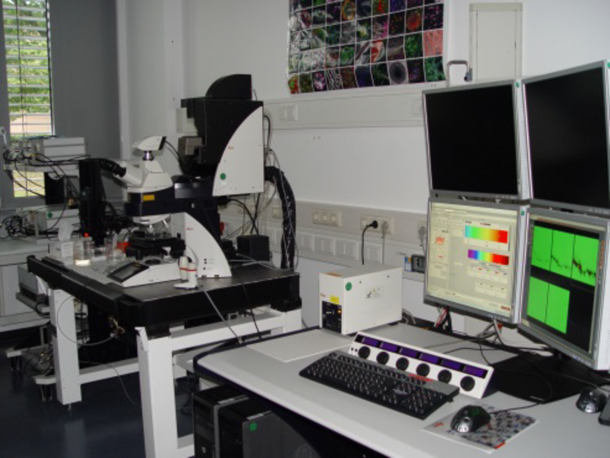
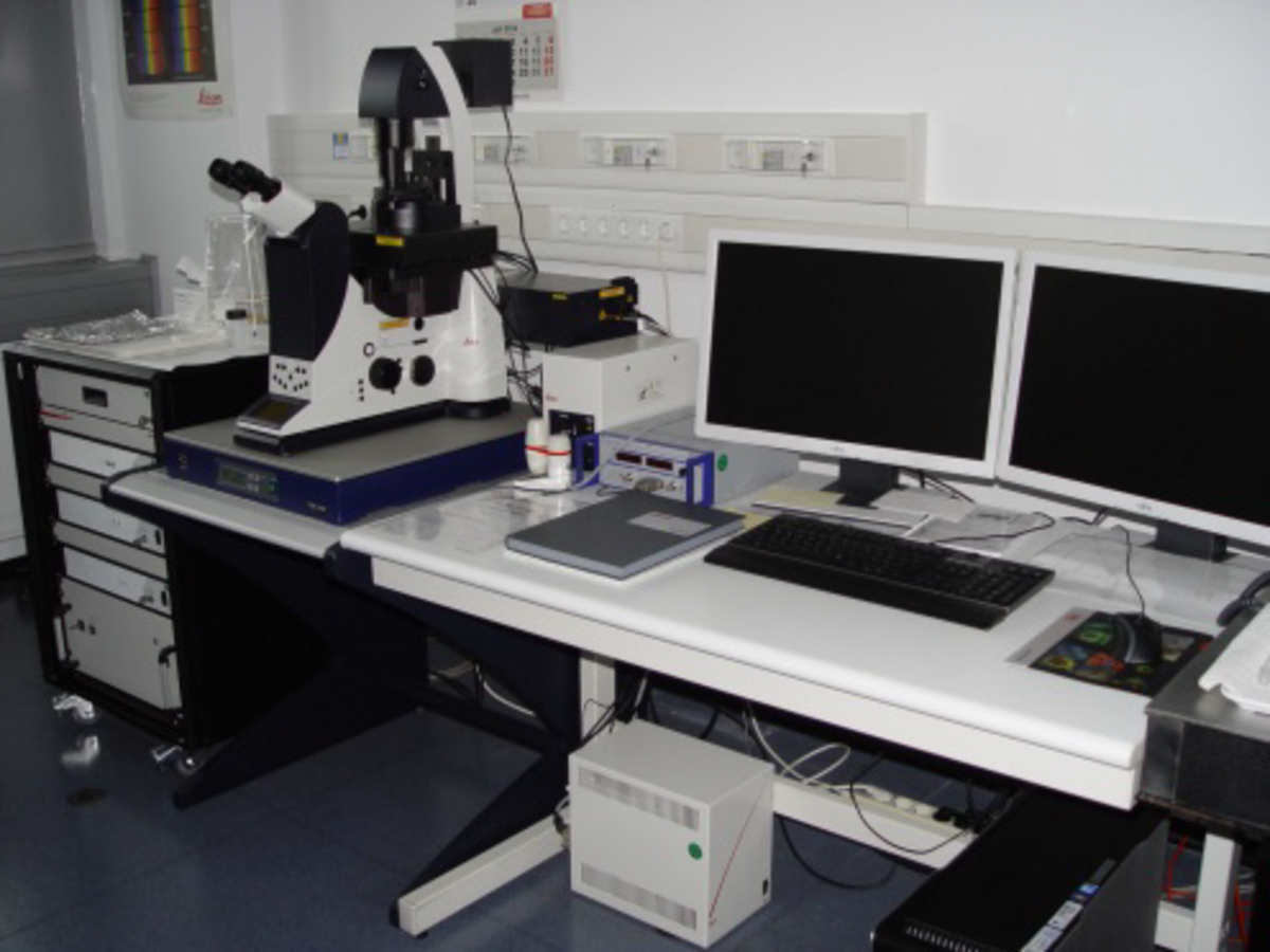
The 3 laser scanning microscopes have been setup and equipped to serve a wide application area, a multitude of sample types and very different research questions. The facility includes the following instruments for laser scanning microscopy and digital image analysis:
- Combined confocal and multi-photon laser scanning microscope, upright and inverted microscope option, continuous visible laser lines at 458, 476, 488, 514, 561, 633 nm, pulsed infrared multi-photon laser 780-900 nm
- Confocal laser scanning microscope, upright microscope, tandem scanner, super continuum light source (white laser) with any line from 470-670 nm Option for fluorescence lifetime imaging (FLIM) with pulsed visible lasers (405, 470, 635 nm) and fluorescence (cross-)correlation spectroscopy/microscopy (FCS)
- GSD nanoscopy (ground state depletion with individual molecule return), laser lines 488, 532, 642 nm, resolution of about 50 nm
- Many options for sample preparation and mounting including cryo-sectioning
- Computer hard and software for digital image analysis: Imaris (visualization) Huygens (deconvolution) ImageJ, J image analyzer (quantification) ConAn (quantification, structural parameters)
The instruments are installed and distributed across 5 laboratories.
Specific features/uniqueness:
- Multi-photon imaging
- White laser excitation
- FLIM, FCS
- Nanoscopy
- Software options for digital image analysis
- 20 years of expertise in analyzing numerous samples from very different habitats
Options and conditions for visiting scientists:
The LSM facility is open to any kind of micro-biological research which is within the wide field of interfacial microbiology, microbial ecology and biotechnology. In addition LSM may be employed for examination of other eukaryotic micro-biological samples as well as technical samples. Applications and proposals for research projects and collaborations will be discussed and considered according to the capacity utilisation and ingenuity of the project.
Unit Cost of use and principles of costing:
Basic costs for running the facility are financed by UFZ. Consumables for in house research are covered by internal funding. Personal costs, travel costs and consumables for external projects have to be paid by the external partners. As an indication for consumables the costs for a fluorochrome / fluorescent probe is in the range of 200–600 euro per unit which however will last for many samples. Other costs for consumables depend on the sample type and mounting.
centre running the infrastructure:
UFZ - Helmholtz Centre for Environmental Research
type of facility:
Laboratory / high-end instrument